
Thoughts on Our Orthodontic Profession Fabio Savastano* International College of Neuromuscular Orthodontics and Gnathology, Italy Citation: Fabio Savastano. “Thoughts on Our Orthodontic Profession”. EC Dental Science 19.10 (2020): 18-19. *Corresponding Author: Fabio Savastano, International College of Neuromuscular Orthodontics and Gnathology, Italy. Received: May 28, 2020; Published: September 10, 2020 30 years have passed since I started my journey in Orthodontics, 25 of which working alone in my office. My wife and my daughter (yes, they are dentist too) share with me my fears and struggles now, comforting me with their presence and energy. Like every other passioned orthodontist, I’m still eager to test my knowledge and update on new trends and issues. I’m constantly in touch with my colleagues and other young dentists that in one way or another have been at my courses or are part of the master’s program where I teach. I gather opinions and watch out to be updated on new tendencies. I got to see TAD’s coming, NiTi and oral scanners just to name a few. I lived through several treatment modalities with aligners and miracle treatments on a weekly basis. Some have disappeared and probably we will never be remembered. Some could very well last and become part of our profession. Defining what a profession is, is not an easy task. It’s a job of course, a craft also. The expertise within should be the result of several components: an orthodontic professional is most certainly driven from a good shovel of vocational beliefs as well as love for the profession itself. A pro should look like a pro, talk like a pro and be wise like a pro. Is that it? Probably not. There is something missing here, and I will try to tell you what it’s about. Let’s take a look at what’s going on in orthodontics in general. First of all, I would like to express my sincere disgust on a new trend that’s taking place all over the world: the superfast treatment 3-3, upper lower or both. Are we really going in the direction of a new sort of “professional” figure, the cosmetic orthodontist? Second, again I would like to express my sincere doubts on the new trend "sagittal first" for the treatment of Class II. This concept represents the milestone of ignorance in orthodontic diagnosis and treatment that crushes crowds of young orthodontists and leads them classify a Class II only as a sagittal problem. Class II is and remains a vertical problem. There is something going on here that is well beyond professionalism. The people that guide all this cannot be so ignorant, I will not accept that, but I can accept that they have lost the most important thing a great leader should have, honesty. They know exactly what they are doing, they are feeding that hungry crowd of what they want most, fast easy and money-making results. Who cares if several patients develop TMD, malocclusion has nothing to do with TMD! (again, I’m disgusted). My last words must focus on something positive, especially after this darn Covid thing. There are orthodontists out there who are leaders in the field and really do their best to guide and change the world. It’s like I’m saying there are some good guys out there that do merit continuous ovations for their effort that appears genuine and separated from the trends I just talked about. You don’t really have to believe in them 100% and embrace their philosophy and interpretation of orthodontics, but I assure you that if you keep your perspective wide and take a look at what they are saying (shouting sometimes) you will be somewhat surprised. I’m going to give you a list of suggested readings and I certainly know that the choice is yours. I know that you will read the list for sure, and I know that some of you will be curious enough to buy a book or click on a website. My greatest effort here is not this suggested reading stuff (even though I wish you would read it), it’s a more deepened message that I hope you get: beware of detractors. Never in these 30 years have I seen such disregard for the Orthodontic Profession. Never in these 30 years I have felt the attack coming from orthodontic companies like in these days. They promote their products through illegitimate orthodontists that have sold their souls for money and promote their pseudoscience with purchased publications. Suggested Readings 1) Jaws, the story of a hidden epidemic, by Sandra Kahn and Paul R. Ehrlich. 2) Buteyko: Close Your Mouth: Buteyko Clinic Handbook for Perfect Health, by Patrick McKeown and Paul Metcalfe. 3) And don’t forget this website on Orthotropics of my friend Mike Mew: https://orthotropics.com/orthotropics-thinkingbeyond- teeth/ |
Neuromuscular Orthodontic Correction of a Class III Patient Fabio Savastano* and Giulia Savastano Abstract Introduction: Patients with a skeletal Class III pattern without reverse overjet may show signs and symptoms of temporomandibular joint (TMJ) overload. Orthodontic correction should be performed while paying particular attention to TMJ preservation. Mandibular tracking and surface electromyography (SEMG) are essential aids for diagnostic and follow-up procedures. Aim: To apply neuromuscular dentistry principles to the orthodontic treatment of skeletal Class III patients with signs and symptoms of TMJ disfunction. Methods: The diagnostic and follow-up procedures were based on the use of mandibular tracking (computerized mandibular scanning), SEMG and transcutaneous electrical nerve stimulation. Results: After treatment and at 10.5 years follow-up, there were no TMD signs or symptoms. Conclusion: Neuromuscular orthodontics is an effective diagnostic procedure that can assist the orthodontist in correcting malocclusions related to TMJ disorders. Keywords: Neuromuscular Orthodontics; Transcutaneous Electrical Nerve Stimulator; Mandibular Posture; Mandibular Kinesiograph; Surface Electromyography |
| Neuromuscular dentistry and the role of the autonomic nervous system: Sphenopalatine ganglion blocks and neuromodulation. An International College of Cranio Mandibular Orthopedics (ICCMO) position paper Ira Shapira, USA The Sphenopalatine Ganglion (SPG) is known to play an integral role in the pathophysiology of a wide variety of orofacial pains involving the jaws, sinuses, eyes and the trigeminal autonomic cephalalgias. It supplies direct parasympathetic innervation to the trigeminal and facial nerves. Sympathetic innervation from the superior sympathetic chain passes thru the SPG to the trigeminal and facial nerves.This paper reviews relevant and significant literature on SPG Blocks and Neuromodulation published in peer reviewed medical and dental journals. Neuromuscular Dentistry employs ULF-TENS to relax musculature and simultaneously provide neuromodulation to the ganglion.Conclusion: The effects of ULF-TENS on the autonomic nervous system acts on the Limbic System and Hypothalamus (H-P-A) to address Axis II issues during neuromuscular dental procedures. It also directly affects the autonomic component of the trigeminal nerve involved in almost all headaches and migraines as well as the Myofascial and Joint disorders of TMD. Shapira, Ira. (2019). Neuromuscular dentistry and the role of the autonomic nervous system: Sphenopalatine ganglion blocks and neuromodulation. An International College of Cranio Mandibular Orthopedics (ICCMO) position paper. Cranio: the journal of craniomandibular practice. 37. 201-206. 10.1080/08869634.2019.1592807. |
Neuromuscular Dentistry: Use and Abuse Savastano, Fabio ** International College of Neuromuscular Orthodontics and Gnathology (ICNOG), Albenga, Italy Full article at https://www.ecronicon.com/ecde/pdf/ECDE-18-00951.pdf |
| Applying neuromuscular techniques in the orthodontic setting. Savastano, Fabio * * International College of Neuromuscular Orthodontics and Gnathology (ICNOG), Albenga, Italy in oral function, hold promise for the treatment of a spectrum of oral disorders. Indeed, such technologies will help patients receive a more targeted level of care. Here we present a case report concerning an 11-year-old boy who underwent orthodontic treatment for recurrent pain of the right temporomandibular joint (TMJ), misaligned teeth, and irregular clicking noises (associated with the right TMJ) during mouth opening. The basic principles of the use of mandibular tracking, surface electromyography, and transcutaneous electrical nerve stimulation (TENS) to diagnose malocclusions and determine the cranio-mandibular relationship are outlined. Case presentation: Pre-treatment status, progress, post-treatment status and 8-year follow-up data are shown. Conclusion: As neuromuscular orthodontics can provide detailed functional analyses through a combination of technologies, the clinician is better placed to evaluate the needs of the patient and deliver treatment. Further deployment of such techniques should, therefore, be encouraged to increase orthodontic health and practice. http://aseestant.ceon.rs/index.php/sejodr/article/download/15529/5731 |
| Effects of upper cervical manipulation on the electromyographic activity of the masticatory muscles and the opening range of motion of the mouth in women with temporomandibular disorder: randomized and blind clinical trial Efeitos da manipulação cervical alta sobre a atividade eletromiográfica dos músculos mastigatórios e amplitude de movimento de abertura da boca em mulheres com disfunção temporomandibular: ensaio clínico randomizado e cego Los efectos de la manipulación cervical en la actividad electromiográfica de los músculos masticatorios y la amplitud del movimiento de apertura de la boca en mujeres con trastorno temporomandibular: un ensayo clínico aleatorizado y ciego Gustavo Luiz Bortolazzo1, Paulo Fernandes Pires2, Almir Vieira Dibai-Filho3, Kelly Cristina dos Santos Berni1, Bruno Mascella Rodrigues4, Delaine Rodrigues-Bigaton5 Mailing address: Delaine Rodrigues Bigaton. Avenida Jaime Pereira, 3701 – CEP: 13403-900 – Piracicaba (SP), Brazil. E-mail: drodrigues@unimep.br – Phone: (19) 97872013 – Presentation: Oct. 2015 – Accepted for publication: Dec. 2015 – Approved by the Ethics Research Committee of the Universidade Metodista de Piracicaba (UNIMEP), under protocol number 01/09 and registered in the Brazilian registry of clinical trials (RBR-4j6xfx). DOI: 10.590/1809-2950/15568322042015 Study performed in the Laboratory of Therapeutic Resources – School of Health Sciences of the Universidade Metodista de Piracicaba (UNIMEP) – Piracicaba (SP), Brazil. 1PhD, Graduate Program in Oral and Dental Biology, Universidade de Campinas (UNICAMP) – Piracicaba (SP), Brazil. 2PhD student, Graduate Program in Human Movement Sciences, Universidade Metodista de Piracicaba (UNIMEP) – Piracicaba (SP), Brazil. 3PhD student, Graduate Program in Rehabilitation and Functional Performance, Universidade de São Paulo (USP) – Ribeirão Preto (SP), Brazil. 4Undergraduate in Physiotherapy (IC), Universidade Metodista de Piracicaba (UNIMEP) – Piracicaba (SP), Brazil. 5Professor, PhD by the Graduate Program in Human Movement Sciences, Universidade Metodista de Piracicaba (UNIMEP) – Piracicaba (SP), Brazil. ABSTRACT | We evaluated the effects of upper cervical manipulation on the surface electromyographic activity (sEMG) of masticatory muscles and range of motion of the opening movement of the mouth in women with temporomandibular disorders (TMD). We evaluated 10 women with myogenic a TMD diagnosis, according to the Research Diagnostic Criteria for Temporomandibular Disorders (RDC/TMD) and divided randomly into an experimental group (EG) n=5, which received upper cervical manipulation, and a placebo group (PG) n=5, which received maneuvers without therapeutic effects. Five interventions were performed in both groups, once a week, with performance of pre-intervention assessments, post-immediate assessments (after 1st intervention) and post-delayed assessments (48 hours after the 5th intervention). The sEMG activity was processed using the root mean square and normalized by the peak value (RMS EMGn). We used for comparison the Student’s 426 t-test and ANOVA two-way repeated measures, adopting as significance the amount of 5%, and the Cohen d for treatment effect size. We found a significant interaction of group vs time (p<0.05) in the RMS EMGn of the left and right temporal muscles at rest, as well as for all masticatory muscles during maximal isometric contraction during jaw elevation and jaw-depression. Treatment effect size, high to moderate, was observed in the EG, especially in the post-delayed assessment. We also observed a significant increase (p<0.05) and a high treatment effect during mouth opening in the EG. The upper cervical manipulation demonstrated a balance of the RMS EMGn of the masticatory muscles and increase the opening range of motion of the mouth in women with myogenic TMD. Keywords | Manipulation, Spinal; Electromyography; Range of Motion, Articular; Temporomandibular Joint Disorders. |
| Does orthodontic proclination of lower incisors in children and adolescents cause gingival recession? Sabine Ruf, DDS, DrMedDent,a Ken Hansen, DDS, OdontDr,b and Hans Pancherz, DDS, OdontDrc Giessen, Germany, and Malmo¨, Sweden In this investigation we sought to assess the effect of orthodontic proclination of lower incisors in children and adolescents with respect to the possible development of gingival recession. Ninety-eight children with a mean 6 SD start-of-treatment age of 12.8 6 1.4 years, treated with the Herbst appliance, were surveyed, for a total of 392 lower incisors. Lateral head films, dental casts and intraoral photographs were analyzed with respect to the degree of orthodontic proclination, crown height, and gingival recession. In all subjects, Herbst treatment resulted in varying degrees of lower-incisor proclination (mean 5 8.9°, range 5 0.5° to 19.5°). In 380 of the surveyed teeth (97%), either no recession developed or preexisting recession remained unchanged during Herbst therapy. In only 12 teeth (3%) did recession develop or preexisting recession deteriorate during treatment. No interrelation was found between the amount of incisor proclination and the development of gingival recession. In conclusion, orthodontic proclination of lower incisors in children and adolescents seems not to result in gingival recession. (Am J Orthod Dentofac Orthop 1998;114:100-6.) |
| The temporomandibular joint in juvenile idiopathic arthritis: frequently used and frequently arthritic. Sarah Ringold*1 and Randy Q Cron2 Address: 1Department of Pediatrics, Division of Rheumatology, Seattle Children's Hospital, Seattle WA, USA and 2Department of Pediatrics, Division of Rheumatology, University of Alabama at Birmingham, Birmingham AL, USA Email: Sarah Ringold* sarah.ringold@seattlechildrens.org; Randy Q Cron - rcron@peds.uab.edu Pediatric Rheumatology 2009, 7:11 Abstract Recent recognition of the markedly high prevalence of temporomandibular joint (TMJ) arthritis in children with juvenile idiopathic arthritis (JIA) coupled with the significant morbidity associated with TMJ damage has prompted increased interest in both the clinical and pathological aspects of TMJ arthritis. This review focuses on the prevalence of TMJ arthritis in JIA, the imaging modalities used to detect TMJ arthritis, and the treatment of TMJ arthritis in children with JIA. |
| Prevalence of clinical signs of intra-articular temporomandibular disorders in children and adolescents A systematic review and meta-analysis Cristhiani Giane da Silva; Camila Pachêco-Pereira, DDS; André Luís Porporatti, DDS, MSc; Maria Gorete Savi, MSc; Marco A. Peres, DDS, MSc, PhD; Carlos Flores-Mir, DDSc, MSc, DSc, RCDC(C); Graziela De Luca Canto, DDS, MSc, PhD Background. The aim of this systematic review and meta-analysis was to assess the prevalence of clinical signs of temporomandibular joint (TMJ) disorders in children and adolescents. Type of Studies Reviewed. The authors selected only studies in which the investigators’ primary objective was to evaluate the prevalence of signs of TMJ disorders according to the international Research Diagnostic Criteria for Temporomandibular Disorders (RDC/TMD) in children and adolescents. The authors performed electronic searches without language restriction in 5 databases. The authors also assessed quality. Results. In this review and meta-analysis, the authors included 11 articles that described studies in which 17,051 participants had been enrolled. The overall prevalence of clinical signs of intra-articular joint disorders was 16% (95% confidence interval [CI], 11.59-19.94; n ¼ 17,051). The prevalence of TMJ sounds (click and crepitation) was 14% (95% CI, 9.67-19.79; n ¼ 11,316). The most prevalent sign was clicking (10.0%; 95% CI, 7.97-12.28; n ¼ 9,665) followed by jaw locking (2.3%; 95% CI, 0.56-5.22; n ¼ 5,735). Conclusions and Practical Implications. One in 6 children and adolescents have clinical signs of TMJ disorders. The results of this systematic research study can alert dentists about the importance of looking for signs of TMD in children and adolescents. Key Words. Evidence-based dentistry; prevalence; children; adolescents; temporomandibular joint disorders. JADA 2015:-(-):--- http://dx.doi.org/10.1016/j.adaj.2015.07.017 |
The role of biochemistry and neurophysiology in the re- education of deglutition Fellus Patrick* Qualified specialist, Former Consultant for the Hospitals of Paris, France Medical and Clinical Archives Review Article ISSN: 2515-1053 Abstract The transition from suction-type deglutition to dentition-type deglutition should spontaneously occur between ages 3 and 4, but the genetic information is not always systematically expressed. The new program must then be engrammed by cortical or sub-cortical pathways. Introduction Suction-type deglutition, the physiological mode for swallowing saliva in the young infant, is a praxis developed in the brainstem in utero. It will remain physiological as long as the primary dentition is not established and mastication has not begun. But beginning at age 4, a new deglutition program is naturally implemented via a subcortical pathway in 60% of children: dentition- type deglutition. By the muscular force it generates, it promotes optimal growth of the maxillae [1]. But endogenous or innate factors can only provide the potential. “They will only be expressed if adequate exogenous conditions occur in a timely manner” (JP Changeux) [2,3]. Dawson Church states that “genes are activated or inactivated by our beliefs, emotions and attitudes.” There are two principal causes for the persistence of suction-type deglutition: ––the child has never had the opportunity to discover this new function ––the child has discovered it, but the limbic system, the gateway to engramming a new program, has not retained it, usually for psychological reasons (immaturity, thumb, pacifier, bottle, food too soft). Most children requiring orthodontic treatment are found in the latter category. The re-education of orofacial functions is consequently a necessity accepted by nearly all practitioners during orthodontic treatment, but it is necessary to properly understand both neuroanatomy and physiology to achieve reproducible and controllable results. Deglutition re-education What strategy to implement? - hope that the normalization of dental arches will lead to a change in function. This is more feasible when wearing a removable retainer. The introduction of an element modifying proprioceptive sensations will automatically lead to a change in the afferent message, and consequently the efferent message [4]. The anatomical part having been modified by treatment, the correct program can install itself naturally, but without control, the new praxis may also remain dysfunctional. However, care must be taken with bonded retainers. If the functional modification occurs, there are no negative consequences, but if the dysfunction remains, the pathological muscular force will be iatrogenic, no longer in the dental alignment but in the supporting bone tissue, which may be the source of subsequent periodontal disease. -use of functional devices, usually at night, lingual envelope-type (NLE) devices intended to modify the lingual posture, Robin device, lingual elevator, Farrell or Bergensen-type gutters. However, the results remain haphazard, as long as the dysfunctional commands are not inhibited, aside from the fact that wearing these devices may be difficult for the child to deal with and rejected by the limbic system. -consultprofessionals,suchasspeechtherapistsorphysiotherapists. The child must initially become aware of what he/she is doing, then the maneuver he/she should be doing, and its repetition should enable automation. Eric Kandel, Nobel Prize in Medicine recipient in 2000 for his work on the transition from short-term to long-term memory, demonstrated inthesecasesanincreaseintheactivityofneurotransmittersinthesynapses involved, but we remain in the domain of short-term memory [5,6]. “Memory is not based on the properties of nerve cells as such, but on the nature of the connections between neurons and how they process the sensory information received”: learning consists of tracing new circuits, and this plasticity occurs either by reconfiguring existing programs or creating new ones (Figure 1 and Table 1). Correspondence to: Fellus Patrick, Qualified specialist, Former Consultant for the Hospitals of Paris, France, E-mail: fellusp@wanadoo.fr Key words: deglutition, short-term memory, procedural memory Received: June 09, 2017; Accepted: June 21, 2017; Published: June 24, 2017 Med Clin Arch, 2017 doi: 10.15761/MCA.1000106 Volume 1(1): 1-3 Patrick F (2017) The role of biochemistry and neurophysiology in the re-education of deglutition Table 1. Reconfiguring existing circuits. 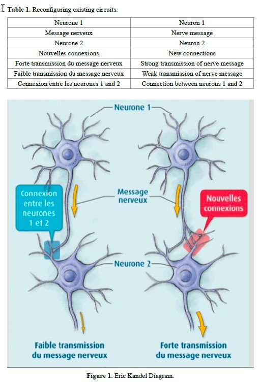 Understanding the transition from short-term memory to long- term memory was clarified by the work of Kandell in Aplysia, by reconstituting a simplified neural circuit: a single sensory neuron connected to a single motor neuron: -A slight stimulus releases neurotransmitters at the synapse, but the nucleus is not involved; this is short-term memory (weekly speech therapy sessions). This information will remain available only briefly. -If the stimuli are repeated soon after, a dialogue occurs between the synapse and the nucleus to activate CREB* and produce a new protein, essential for the transition to long-term memory. This new CPEB** protein present in the synapse will function as a prion and permanently ensure the transmission of the message. *CREB (Cyclic AMP Response Element-Binding Protein), a protein that activates the genes responsible for long-term memory. CREB 1 is the activator and CREB is the 2 inhibitor. **CPEB: (Cytoplasmic Polyadenylation Element-Binding Protein), a transcriptional regulatory protein in the synapse which contributes to the stabilization of long-term memory. Creating new circuits A highly emotional state can short-circuit normal constraints and produce a sufficient quantity of MAP-kinase* molecules, which will be sent to the nucleus to inactivate CREB-2** molecules and facilitate CREB-1** activation and direct imprinting of this experience in long- term memory *MAP-kinase kinase: acts in conjunction with protein kinase A, to initiate long-term memorization. - FroggyMouth is a device worn a rather short time, 15 minutes a day while watching television (a reward recognized by the limbic system), which will force the child to discover a new deglutition method via the sub-cortical pathway; thus, not by stimulating neurotransmitter activity, but by creating new synapses. In fact, by no longer being able to close the lips, the child will be unable to swallow by suction, aspirating between the anterior and posterior mouth, triggering an abrupt and immediate reaction in the brainstem: find a new deglutition program. The simultaneous contraction of the levator muscles of the mandible in a stable and comfortable dental occlusion with those of the soft palate and styloglossus will allow a peristaltic movement of the tongue (provided that the anatomical environment is compatible) and disconnect the tongue-lips synkinesis. This new deglutition program will be immediately integrated into long-term memory by the creation of a new neural circuit. But this is only the first step, which is necessary but not sufficient for the transition to automation. Automation The child will then have two programs for swallowing saliva, and just as on a computer when there are two programs, it is the activation of one or another icon that initiates its execution (Figures 2 and 3). The therapist should therefore monitor the resting posture for relaxation of the perioral muscles and dental occlusion upon swallowing. Control by the trigeminal nerve, solicited at this step, will replace control by the facial nerve and inhibit the role of the latter. This necessary inhibition of the faulty circuit is fundamental to automation of the correct program. Only FroggyMouth allows this double action. The trigeminal nerve, which also controls the respiration centers in the pontine tegmentum through its sensory nucleus, will promote restoration of nasal respiration, allowing the tongue to adopt a high posture posteriorly (the lingual dome). “This dual posterior and occlusal functional necessity of the tongue, too often forgotten by re-educators of orofacial function, is likely one of the causes of the too-frequent failures of re-education” (Delaire) [7]. Similarly, contraction of the tensor tympani muscle, innervated by the trigeminal nerve, will allow ventilation of the middle ear by dilating the Eustachian tube, decreasing serous otitis problems Control may be transferred to the parents, who need to observe lip posture 5 times/ day, and who then congratulate or correct the child (Figures 4 to 8). These two actions are not similar, involving cortico-cortical circuits that traverse the basal ganglia and cortico-cortical circuits that traverse the cerebellum [8]. Wearing FroggyMouth starting at age 3 has no contraindications. Conclusion Early normalization of orofacial functions, regardless of the technique chosen, will enable a three-step preventive approach: Med Clin Arch, 2017 doi: 10.15761/MCA.1000106 Volume 1(1): 2-3 Patrick F (2017) The role of biochemistry and neurophysiology in the re-education of deglutition 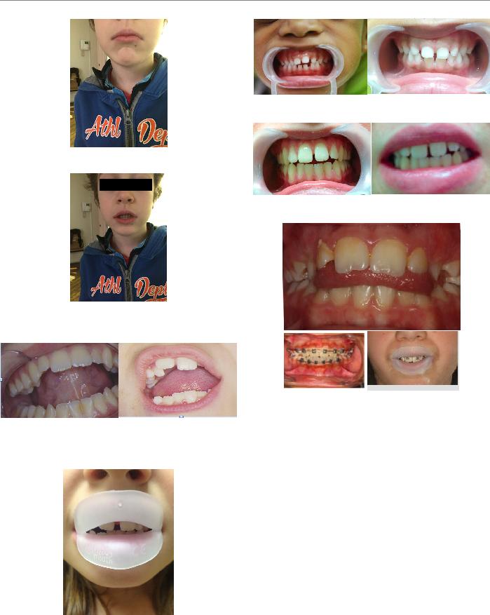 Figure 2. The suction-type deglutition icon is activated by the facial nerve: “My lips are contracted, my teeth do not touch.” Figure 3. The dentition-type deglutition icon: “my lips are relaxed, my molars are in occlusion” is activated by the trigeminal nerve, whichallows not onlymolar occlusion but also the protection of the tongue from biting, due to the abundance of trigeminal nerve endings in its epitheliallining. Figure 4. Contrary to what speech therapists suggest, attention needs to be paid not to the tip of the tongue, but rather to its posterior portion. Obsessedwith the sensorysearch for the retro-incisive papilla, the child risks raising the tip of the tongue, leading to lowering of the posterior portion, which will prevent the involvement of the styloglossus, the levator muscle of the lingual dome. Figure 5. Worn for 15 min daily while watching television, FroggyMouth allows relaxation of all anterior facial muscles. Figure 6. Spontaneous improvement of an incisor gap after wearing Froggy Mouth for one month. Figure 7. Resumption of treatment with only Froggy Mouth after repetition of a previous orthodontic treatment. Figure 8. Case treated by Gérard Altounian. -prevent deformities from appearing, -if they occur, prevent them from worsening, References 1.Patrick F (2003) Orthodontie précoce en denture temporaire Cdp. 2.Jean-Pierre C (1983) L’homme neuronal Fayard. 3.Gérard C (2010) Les oralités humaines Doin. 4.Gérard C (2015) Oralité du fœtus Sauramps Médical. 5.Arthur G (1996) Neurosciences. Piccin. 6.Eric K (2011) A la recherche de la mémoire. Odile Jacob. 7.Patrick F, Waddah S, Lalauze-Pol R (2016) De la dysfonction à la dysmorphose. Apport de Froggy mouth. Edition Orthopolis. 8.Fournier M, Girard M (2013) Acquisition and sustainment of automatic reflexes in maxillofacial rehabilitation. Orthod Fr 84: 287-294. [Crossref] Copyright: ©2017 Patrick F. This is an open-access article distributed under the terms of the Creative Commons Attribution License, which permits unrestricted use, distribution, and reproduction in any medium, provided the original author and source are credited. Med Clin Arch, 2017 doi: 10.15761/MCA.1000106 Volume 1(1): 3-3 |
MASTICATORY FUNCTION IN TEMPOROMANDIBULAR DYSFUNCTION PATIENTS: ELECTROMYOGRAPHIC EVALUATION FUNÇÃO MASTIGATÓRIA EM PACIENTES COM DISFUNÇÃO TEMPOROMANDIBULAR: AVALIAÇAO ELETROMIOGRÁFICA Giédre BERRETIN-FELIX1, Katia Flores GENARO2, Inge Elly Kiemle TRINDADE3, Alceu Sergio TRINDADE JÚNIOR3 1- DDS, MSc, PhD, Professor, Department of Speech, Language and Audiology, Bauru Dental School, University of Sao Paulo (FOB-USP), Bauru, SP, Brazil. 2- DDS, MSc, PhD, Associate Professor, Department of Speech, Language and Audiology, Bauru Dental School, University of Sao Paulo (FOB-USP), Bauru, SP, Brazil. 3- DDS, MSc, PhD, Associate Professor, Department of Biological Sciences, Bauru Dental School; University of Sao Paulo (FOB-USP), Bauru, SP, Brazil. Corresponding address: Department of Speech Pathology and Audiology, University of São Paulo - Al. Dr. Octávio P. Brisolla, 9-75, 17012- 901 - Bauru - SP - Brazil. - E-mail: gfelix@fob.usp.br Received: February 25, 2005 - Modification: May 10, 2005 - Accepted: June 30, 2005- J Appl Oral Sci. 2005;13(4):360-5 Temporomandibular dysfunction (TMD) is a complex disturbance that involves the masticatory muscles and/or temporomandibular joint, causing damage to the masticatory function. This study evaluated the electromyographic activity of the masseter muscle during habitual mastication of bread, apple, banana, cashew nut and paraffin film (Parafilm M) in 25 adult subjects, of both gender, with TMD. The results were compared to those of a control group, composed of 15 adult subjects, of both sexes, free of signs and/or symptoms of TMD. The MYO-TRONICS Inc., K6-I computer software was used for electromyographic processing and analyzed the following parameters: duration of the act, duration of the masticatory cycle and number of cycles. No significant differences were found between subjects in the control group and individuals with TMD as to duration of the masticatory act and of the masticatory cycle, considering all materials used for mastication. The duration of the masticatory act and cycle was longer during mastication of paraffin film in both groups. The number of masticatory cycles was higher for mastication of apple in comparison to mastication of banana, in both groups. It can be concluded that the consistency of foods influences the duration parameters of the act, duration of the cycle and the number of masticatory cycles, and the behavior of the masticatory muscles in individuals with TMD during habitual mastication is similar to that verified in individuals without TMD. Uniterms: Mastication; Electromyography; Food; Temporomandibular joint disorders. |
Go to content
Back to content